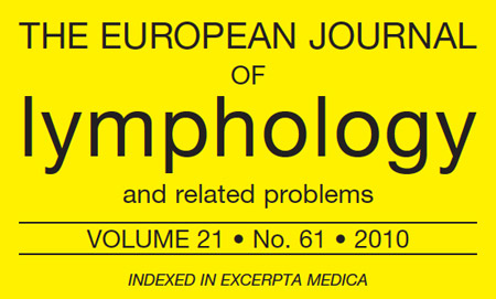
Here it is a full publication from prof. Boccardo
- This pictures show the use of LY.M.P.H.A. technique to prevent extremity lymphedema following cancer treatment.
- Despite the numerous efforts to reduce complication rates after ilioinguinal lymphadenectomy, morbidity following inguinofemoral lymphadenectomy can be substantial, and the treatment and prevention of lymphedema remains a true challenge.
- This picture shows Ly.M.P.H.A. technique at the left axilla after lymphnodal dissection for breast cancer. Following our experience for arm lymphedema, we assessed the feasibility of performing Ly.M.P.H.A. technique after inguinofemoral lymph node completion and the possible benefit of it for preventing lower limb lymphedema.
- The technique is shown in this picture: multiple lymphatic-venous anastomoses are performed after inguinofemoral lymph node completion according to Ly.M.P.H.A. technique. Lower limb lymphatics are colored in blue thanks to the BPV previously injected into the thigh. In this case, the superficial epigastric vein was used for lymphatic-venous anastomoses.
- 11 patients with vulvar cancer and 16 patients with melanoma of the trunk requiring inguinofemoral lymphadenectomy underwent lymph node dissection and Ly.M.P.H.A. technique.
- Patients (R right; L left; BIL bilateral) : These tables show the number of lymphatics anastomosed (4 collectors averagely), the vein used (Medial Accessory, Superficial Circumflex, Superficial Epigastric or Lateral Accessory) and the time taken to perform LYMPHA technique (27 minutes in average).
- This patient underwent bilateral groin lymphnodal dissection plus Ly.M.P.H.A. for the treatment of a vulvar cancer; the follow-up is at 3 ys. Ultrasonography demonstrated long term patency of MLVA.
- Even with the videoendoscopic approach for inguinofemoral lymphadenectomy, it is possible to prevent lymphatic complications using the blue dye which permits to visualize lymphatics and ligate them with metallic clips and performing multiple lymphatic-venous anastomoses after nodal dissection according to LYMPHA technique.
- The LYMPHA technique appears feasible, safe and effective for the prevention of both upper and lower limb lymphedema, thereby improving the patient’s quality of life and decreasing healthcare costs. The key of success is based on an interdisciplinary approach! Thank you very much for your kind attention!
In collaboration with: Sara Dessalvi, Lidia Molinari, Stefano Spinaci, Corrado C.Campisi, Corradino Campisi

